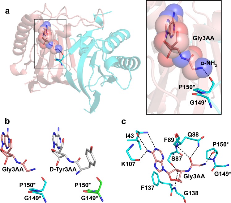
Fig 3. Glycine binding mode in the chiral proofreading site of DTD.
(a) Co-crystal structure of PfDTD with Gly3AA showing the capture of the ligand. (b) Comparison of PfDTD+Gly3AA complex with PfDTD+D-Tyr3AA complex (PDB id: 4NBI) showing the flipped orientation of the α-NH2 group of Gly3AA. (c) Network of interactions of Gly3AA with active site residues of PfDTD. Residues indicated by * are from the dimeric counterpart.

