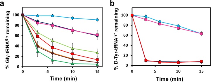
Fig 5. EF-Tu confers protection on Gly-tRNAGly.
(a) Deacylation of Gly-tRNAGly in the presence of unactivated EF-Tu (dark blue circle), activated EF-Tu (light blue circle), unactivated EF-Tu and 5 nM EcDTD (red square), activated EF-Tu and 5 nM EcDTD (pink square), unactivated EF-Tu and 10 nM EcDTD (dark green triangle), activated EF-Tu & 10 nM EcDTD (light green triangle), unactivated EF-Tu and 20 nM EcDTD (dark brown diamond), and activated EF-Tu and 20 nM EcDTD (light brown diamond). (b) Deacylation of D-Tyr-tRNATyr in the presence of unactivated EF-Tu (light blue circle), activated EF-Tu (pink square), unactivated EF-Tu and 5 nM EcDTD (dark blue circle), activated EF-Tu and 5 nM EcDTD (red square). Error bars indicate one standard deviation from the mean. The underlying data can be found in S1 Data.

