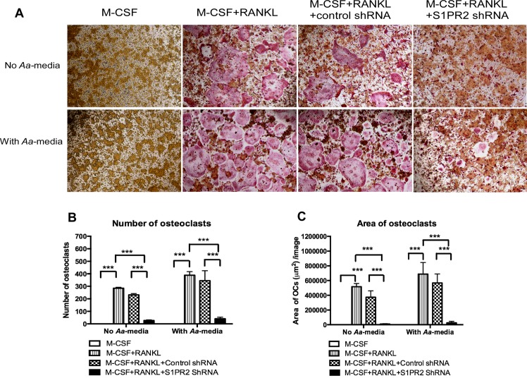
Fig 3. Knockdown of S1PR2 suppressed osteoclastogenesis in BM cells induced by RANKL.
BM cells were uninfected, infected with a S1PR2 shRNA lentivirus, or infected with a control shRNA lentivirus (moi 20), and co-cultured with M-CSF and RANKL as described in Methods. A control group of cells were cultured only with M-CSF. BM cells were either untreated or treated with A. actinomycetemcomitans-stimulated media (Aa-media) for 24 h. (A) Representative images show TRAP-stained cells with and without Aa-media stimulation. Pictures were taken at 100x magnification. (B) Number of TRAP+ multinucleated (more than 3 nuclei) osteoclasts/well (96-well) and (C) Total areas of osteoclasts/image were quantified. The data are representatives from three separate experiments (n = 3, ***P<0.001).

