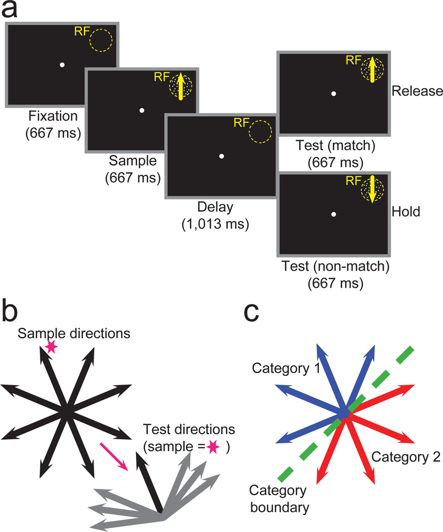
Figure 1. DMS and DMC tasks.
(a) Monkeys performed a delayed matching task and indicated (by releasing a lever) whether sample and test stimuli, separated by a memory delay, were identical (DMS) or category (DMC) matches. If the sample and test stimuli weren’t matches, the monkey was required to continue holding the lever throughout the test period and a second delay period until a second, matching test stimulus appeared. Stimuli were shown in a neuron’s RF. (b) Monkeys viewed eight motion directions as sample stimuli during DMS task recordings (left). Test stimuli were either identical matches or 45°, 60°, or 75° from the sample stimulus. As an example, the possible non-matching test directions are shown for the sample direction indicated by the magenta star. (c) Monkeys grouped the same eight motion directions used as sample directions during the DMS task into two categories (corresponding to the red and blue arrows) separated by a learned category boundary (dashed green line). Test and sample stimuli sets were identical.

