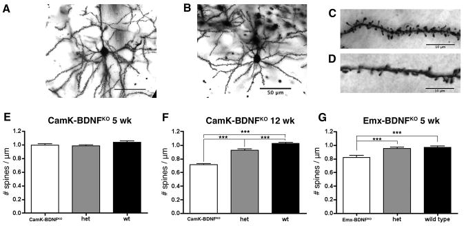
Fig. 6.
Spine density in visual cortex is reduced in CaMK-BDNFKO mice at 12 weeks, but not at 5 weeks. A) and B) Typical layer II/III pyramidal cell in visual cortex V1M of wild type (A) and CaMK-BDNFKO mice (B). C) and D) Spines on basal dendrites of layer II/III pyramidal cells in wild type (C) and CaMK-BDNFKO mice (D). E) Spine density is unaltered in 5 week old CaMK-BDNFKO mice compared to wild type controls (n = 70 dendritic segments on 35 neurons from 5 animals of each genotype). F) Spine density is reduced in 12 week old CaMK-BDNFKO mice compared to wild type controls (n = 70 dendritic segments on 35 neurons from 5 animals of each genotype). G) Spine density is reduced in 5 week old Emx-BDNFKO mice compared to wild type controls (n = 56 dendritic segments on 28 neurons from 4 animals of each genotype). *** p< 0.001.

