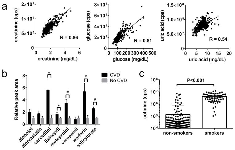
Figure 1. Examination of select metabolomics data in relation to CRIC phenotyping.
(a) Correlation between LC-MS (y-axis) and spectrophotometric measures (x-axis) of creatinine, glucose, and uric acid. (b) Mean medication levels in individuals with (n=166) or without (n=234) self-reported CVD at baseline. Bars represent SEM. *p < 0.05, #p < 0.001. (c) Distribution of plasma cotinine levels in self-reported non-smokers and smokers; note y-axis is log scale.

