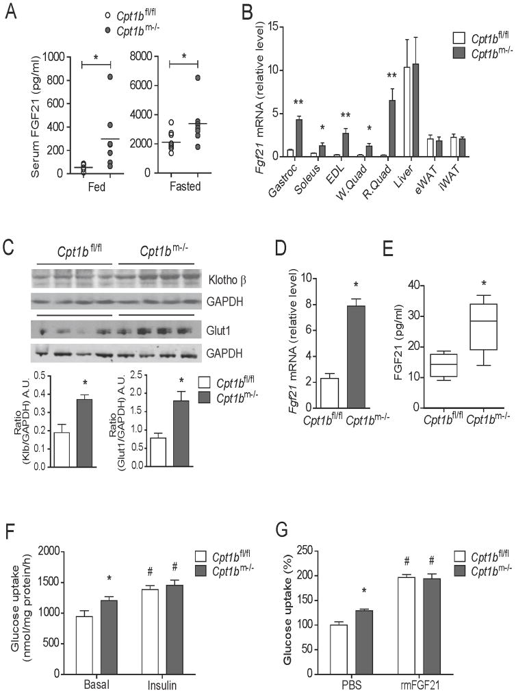
Figure 1. Muscle-derived FGF21 enhances glucose uptake in skeletal muscle in Cpt1bm−/− mice.
(A) Serum FGF21 concentration in 4 month old Cpt1bm−/− and Cpt1bfl/fl mice fed and fasted overnight (n=7–9 per group). (B) Fgf21 gene expression in muscle tissue such as gastrocnemius (Gastroc), soleus, extensor digitorium longus (EDL), white and red quads (W.Quad and R.Quad), and other tissue such as liver, epididymal and inguinal white adipose tissue (eWAT and iWAT) from 3–5 month old Cpt1bm−/− and Cpt1bfl/fl mice (n=5–8). (C) Immunoblot analysis showing Klotho β and Glut1 protein abundance in gastrocnemius muscle from 4 month old Cpt1bm−/− and Cpt1bfl/fl mice. GAPDH used as a loading control. Image J software was used for densitometry quantification of the immunoblots. Results shown are representative of three separate experiments (n=3–4 per group). (D) Fgf21 gene expression primary myotubes established from Cpt1bm−/− and Cpt1bfl/fl mice. (E) Secreted FGF21 over 24h in culture media from primary myotubes initiated from Cpt1bm−/− and Cpt1bfl/fl mice. (F) Basal and insulin-stimulated [3H]-2-deoxyglucose uptake in primary myotubes from Cpt1bm−/− and Cpt1bfl/fl mice. (G) Primary myotubes from Cpt1bm−/− and Cpt1bfl/fl mice (3–5 months of age) were pre-treated with vehicle (PBS) or rmFGF21 (250 ng/ml, 16h) and then [3H]-2-deoxyglucose uptake was measured. Results shown are representative of four independent experiments. All data are presented as means ± s.e.m., *P < 0.05 and **P < 0.01 between Cpt1bm−/− and Cpt1bfl/fl mice, and #P < 0.05 between vehicle versus insulin or rmFGF21 treatments.

