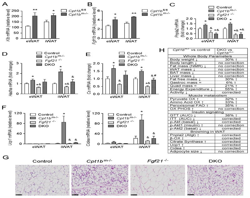
Figure 7. Muscle-derived FGF21 enhances browning in white adipose tissue in Cpt1bm−/− mice.
(A–B) Gene expression of Klb (A) and Fgfr1b (B) in epididymal and inguinal white adipose tissue (eWAT and iWAT) (n=5–7 per group). All data are presented as means ± s.e.m., *P < 0.05, **P < 0.005 and higher significance. (C–E) qRT-PCR analysis of Expression of a lipolytic gene (Pnpla2) (C), mitochondrial FAO genes (Hadha, Cs) (D–E), and beige adipocyte marker genes (Ucp1, Cidea) in WAT (F). (G) H&E staining of iWAT. Scale bar indicates 100 μm. (H) Summary of phenotype in Cpt1bm−/− mice compared to control mice and the reverse phenotype in DKO mice. Tissue from control (Fgf21+/+ Cpt1bfl/fl), Cpt1bm−/− (Fgf21+/+ Cpt1bm−/−), Fgf21−/− (Fgf21−/− Cpt1bfl/fl) and DKO (Fgf21−/− Cpt1bm−/−) mice were used for all qRT-PCR analysis (n=5–7 per group). All data are presented as means ± s.e.m., *P < 0.05 significance compared to control mice, &P < 0.05 significance compared to Cpt1bm−/− mice and #P < 0.05 significance compared to Fgf21−/− mice.

