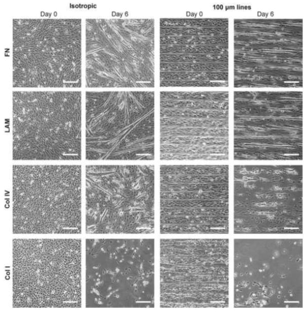
Figure 1.
Phase contrast images of C2C12 cells differentiated on FN, LAM, Col IV, and Col I. Samples were differentiated on isotropically coated coverslips and 100×20 micropatterns. After 6 days of differentiation, cells begin to delaminate from μCP lines of Col IV and Col I and isotropically coated Col I. However, myotubes were able to form on both μCP or isotropically coated LAM and FN. Scale bars 200 μm.

