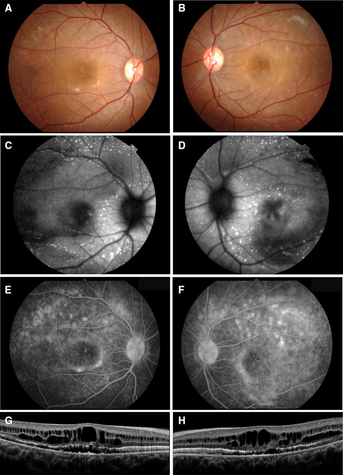
Fig. 1.
Fundus photographs, autofluorescence images, fluorescein angiograms, and SD-OCT images from patient with autosomal recessive bestrophinopathy (ARB) (proband, II-1). Fundus photographs (a, b), autofluorescence images (c, d), fluorescein angiograms (e, f), and SD-OCT images (g, h) are shown. Results from the right eye (a, c, e, g) and left eye (b, d, f, h) are shown. Fundus photograph shows cystoid macular lesions and multiple yellowish deposits throughout the posterior pole of both eyes. FAF images show multiple hyper-autofluorescent regions in the peripheral retina of both eyes. FAF images also show a hypo-autofluorescent lesion in the macular of both eyes. Fluorescein angiograms show widespread patchy hyper-fluorescence. The SD-OCT images show cystoid macular changes and shallow serous retinal detachments in both eyes. There is also a thickening and hyper-reflectivity at the areas corresponding to the ellipsoid and interdigitation zones

