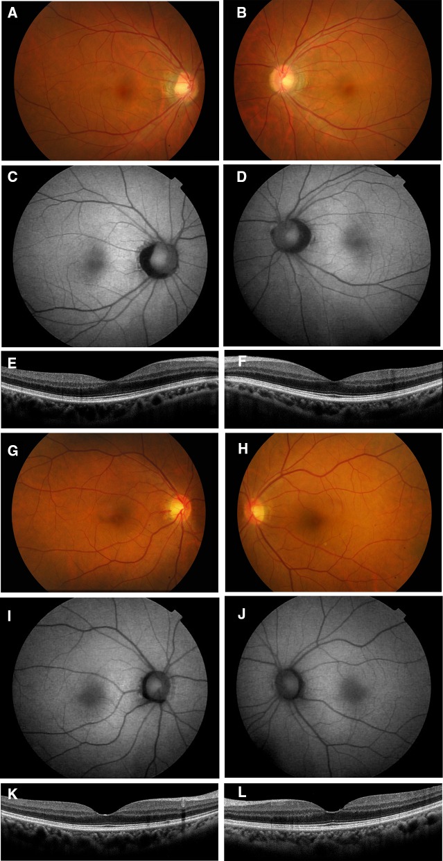
Fig. 6.
Fundus photographs, fundus autofluorescence image, and SD-OCT images from the parents of the proband. Fundus photographs (a, b, g, h), autofluorescence (c, d, i, j), and SD-OCT images (e, f, k, l) are shown. Results from the father (a–f) and mother (g–l) are shown. Fundus appearance, FAF, and SD-OCT of the proband’s parents are normal

