Figure 2.

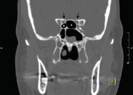
Variations of the posterior paranasal sinuses and related structures. Bilateral Onodi cell (white star), left dehiscent internal carotid artery (ICA) (white arrow). Optic nerve in relation to Onodi cell (black arrows).

Variations of the posterior paranasal sinuses and related structures. Bilateral Onodi cell (white star), left dehiscent internal carotid artery (ICA) (white arrow). Optic nerve in relation to Onodi cell (black arrows).