
Figure 4.
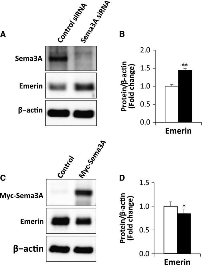
Sema3A regulated protein expression of emerin. (A) Myoblasts were transfected with Sema3A or control siRNA. After 2 days of transfection in GM, cells were lysed and lysates analyzed by western blotting for protein expression of Sema3A and emerin. β‐actin was used as an internal control. (B) Representative western blots showing emerin protein expression in myoblasts transfected with Sema3A or control siRNA (A). The results are expressed as values relative to β‐actin expression. Data are means ± S.E. *P < 0.05, **P < 0.01 vs. control siRNA. (C) Myoblasts were transfected with Myc‐Sema3A or control vector. After 2 days of transfection in GM, cells were lysed and subjected to western blotting. Protein expression of Myc‐Sema3A and emerin were identified using Myc and emerin antibodies, respectively. β‐actin was used as an internal control. (D) Representative western blots showing protein expression of emerin in myoblasts transfected with Myc‐Sema3A or control vector (Fig. 3A). Results are expressed as values relative to those of β‐actin expression. The data are means ± S.E. *P < 0.05, **P < 0.01 vs. control vector.