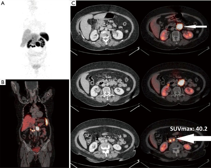
Figure 5.
A 67-year-old female presented with metastatic mesenteric lymph node (CUP-NET). Ga-68 DOTANOC PET/CT scan revealed at least three avid nodular lesions in third part of duodenum (thicker arrow) (site of primary) with lymph nodal metastases (thin arrow). No surgery was performed in this patient. (A) PET MIP image; (B) fused PET/CT coronal image; (C) fused PET/CT trans-axial images. NET, neuroendocrine tumour; PET/CT, positron emission tomography/computed tomography; MIP, maximal intensity projection.

