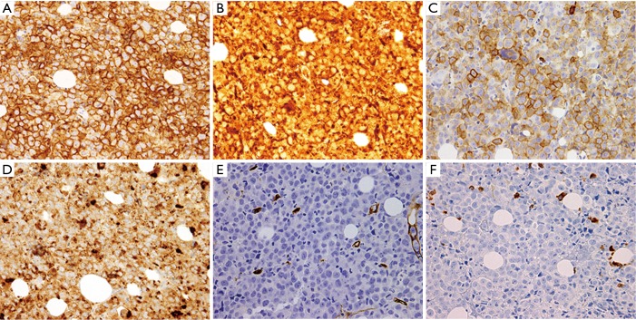
Figure 4.
Immunohistochemistry (IHC) of bone marrow biopsy showing: (A) CD33 diffusely positive staining (magnification, ×400); (B) CD68 diffusely positive staining (magnification, ×400); (C) CD117 positive staining in numerous cells (magnification, ×400); (D) lysozyme diffusely positive staining (magnification, ×400); (E) CD34 absent staining in the tumor cells (magnification, ×400); (F) myeloperoxidase (MPO) is negative in the tumor cells (magnification, ×400).

