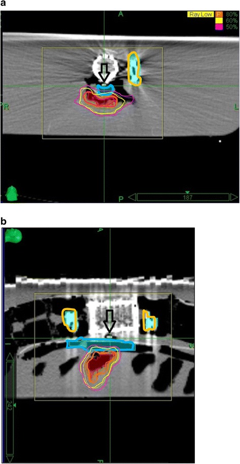
Fig. 2.

a and b. TLD (Thermoluminescent dosimeter) placement in the AIAC model. Figure 2A shows the axial and 2B shows the sagittal views in treatment position. The arrow shows the TLD behind the vertebral body, anterior to the spinal cord. The blue-outlined and orange-outlined structures represent the spinal cord and rods, respectively. The red structure represents the target volume. The orange, yellow and pink lines represent 80 %, 60 %, and 50 % isodoses respectively
