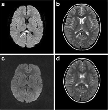
Fig. 1.

MRI findings of case 1. Diffusion-weighted image (a) and T2-weighted image (b) on the day of admission showed a focal high-signal lesion in the splenium of the corpus callosum (arrow). On the seventh day after admission, the splenial lesion had completely disappeared on diffusion-weighted image (c) and T2-weighted image (d)
