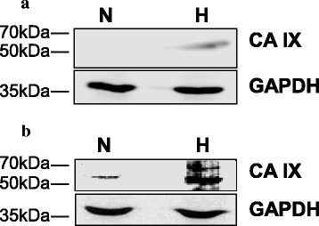
Fig. 1.

Western blot analysis of CA IX in LNCaP and PC-3 cells, under normoxia and hypoxia. Low protein expression is detected in LNCaP (a) and PC-3 (b) cells under normoxia (N). Under hypoxia (H) the staining becomes evident and is stronger in PC-3 than LNCaP cells. GAPDH is used as control for protein loading
