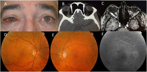
Fig. 1.

a Initial photograph showing tortuous and dilated episcleral vessels of the patient’s right eye. b CT shows an enlarged right superior ophthalmic vein. c MRA suggested a reduction of blood flow, which led to the suspicion of cavernous sinus-dural arteriovenous fistula. d Fundus photography of the right eye with venous congestion of the retinal veins and macular subretinal fluid. e The normal left eye. f Fluorescein angiography without clear leakage in the area of the serous detachment
