Figure 1.
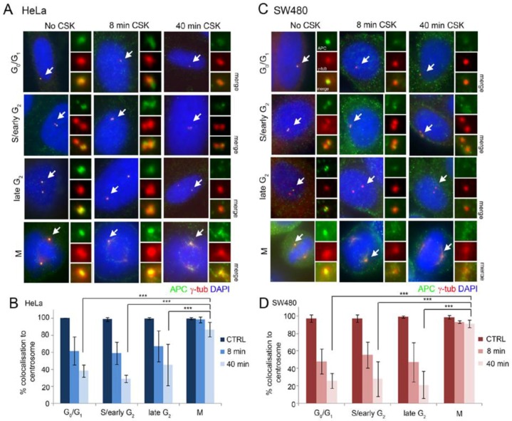
APC is less retained at centrosomes in interphase than mitotic cells. Immunofluorescence micrographs of APC at centrosomes after CSK detergent buffer washout in (A) HeLa cells (express full-length APC (APC-FL)), and (C) SW480 (express mutant APC1-1337). Cells were stained with antibodies against APC (Ab7 mAb, green) and γ-tubulin (red) to detect centrosomes (white arrows). The localization of retained APC was scored at different stages of the cell cycle, which were classified according to centrosome number and position (Go/G1 = 1 centrosome, S/G2 = 2 centrosomes, late G2 = separated centrosomes that have not yet matured, M = divided centrosome with increased γ-tubulin at the centrosome). APC localization was visually scored by microscopy after 8 min and 40 min detergent washout, and compared to cells with no detergent treatment (No CSK) (n = 300). (B and D) Scores pooled from three experiments were normalized to indicate the percentage of centrosome localization based on ability to clearly visualize APC by direct fluorescence microscopy with adjustment of the Z-axis focus (mean ± SD) (***, p < 0.001). Additional cell images of APC in mitotic cells are shown in Figure S1.

