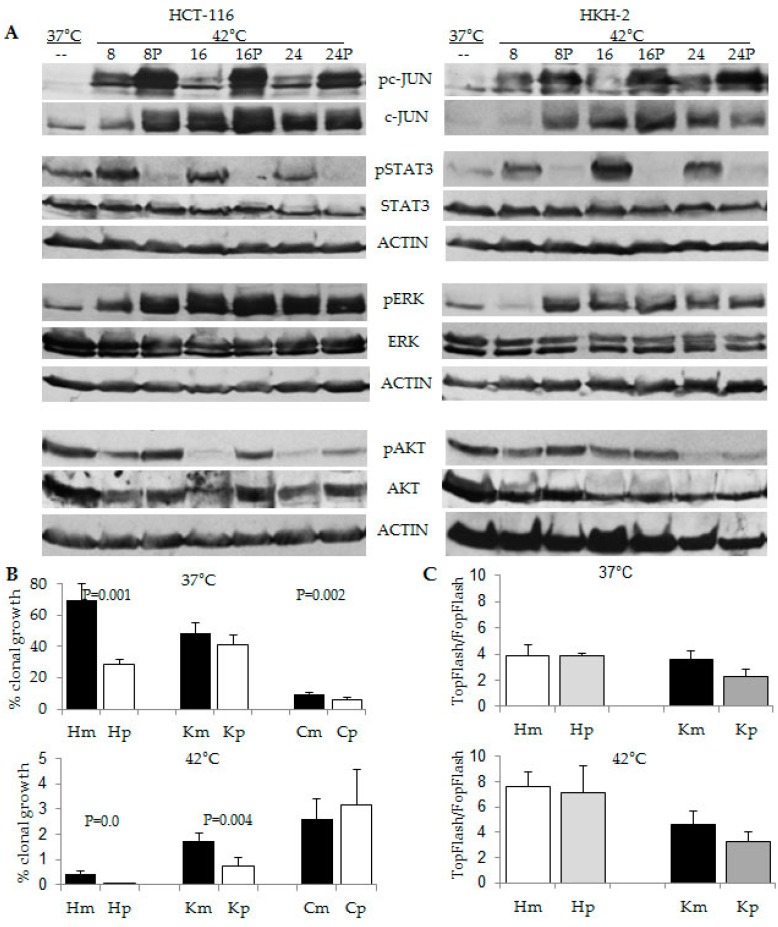
Figure 7.
Combined effect of hyperthermia and propolis on CRC cells. (A) Representative Western blot analyses with total cell lysates after exposure of HCT-116 and HKH2 cells to 37 °C or 42 °C, for 8, 16, or 24 h in absence or presence of 100 μg/ml propolis (P); (B) Clonal growth assays of HCT-116 (H), HKH2 (K) and CDC841CoN (C) cells were performed as described in the legend of Figure 1. Cells were exposed simultaneously for 24 h to 37 °C or 42 °C and to mock (m) treatment or propolis (p) at 100 μg/mL. Results are the mean from minimum of four clonal growth assays with triplicate samples per treatment. Only statistically significant different values are indicated; (C) WNT/beta-catenin transcriptional activity was measured with the luciferase reporter pair TOP-Flash and FOP-Flash as described in Materials and Methods. Mock and propolis treatment were performed for 16 h as in (B).

