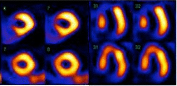
Fig. 2.

Perfusion defect on myocardial perfusion scintigraphy. Myocardial perfusion scintigraphy with Tc-99 m Sestamibi: short-axis and horizontal long-axis images during stress (upper rows) and rest (lower rows). A severe reversible perfusion defect is seen in the anteroseptal area of the left ventricle
