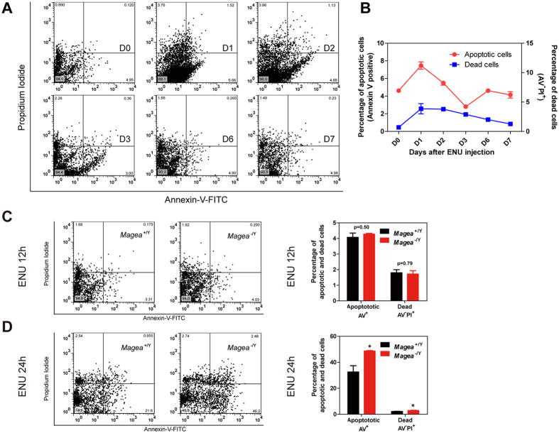
Figure 4. Magea proteins prevented an excessive apoptotic response in germ cells in an acute manner after ENU-induced genotoxic stress.
(A) Time course flow cytometry analysis of testicular apoptosis in C57BL/6J mice after exposure to ENU. The results are shown at several time points (in days) as indicated. (B) Quantification of apoptosis after exposure to ENU for 1 week (n = 4 for each time point). (C,D) Detection of testicular apoptosis by flow cytometry analysis at 12 hours and 24 hours after exposure to ENU (60 mg/kg) (12 hours, n = 4 for wild type and n = 5 for Magea−/Y; 24 hours, n = 6 for wild type and n = 4 for Magea−/Y). *p < 0.05.

