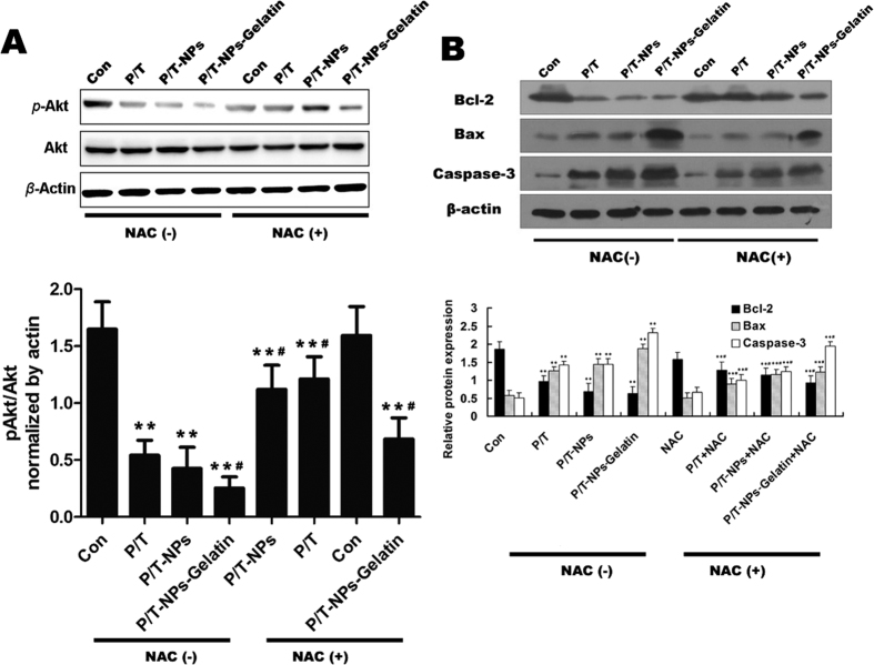
Figure 10.
(A) the expression of p-Akt and Akt in tumors extracted from different groups of mice. Semi-quantification of the gel image, normalized to β-actin control. Each data point represents the mean ± SD; **represents p < 0.01 versus control. #represents p < 0.05 versus the corresponding group without the presence of N-acetyl cysteine (NAC). (B) The expression of Bcl-2, Bax and Caspase-3 in tumors extracted from different groups of mice. Semi-quantification of the gel image, normalized to β-actin control. Each data point represents the mean ± SD from three independent experiments; **represents p < 0.01 versus control. #represents p < 0.05 versus the corresponding group without the presence of NAC.

