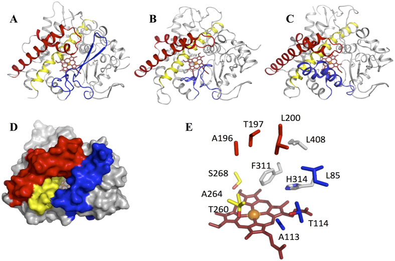
Figure 7. The crystal structure of CYP144A1-TRV.
Panels (A,D) Ribbon and surface representations, respectively, of the Mtb P450 CYP144A1-TRV. The heme, BC-loop, FG-helices and I-helix are coloured red, blue, dark red and yellow, respectively. Panels (B,C) Ribbon structure representations of Mycobacterium smegmatis CYP142A2 (PDB 2YOO) and the putative orsellinic acid oxidase P450 CalO2 (PDB 3BUJ) using similar colour coding as panel (A). The orientation of both structures is similar to that of CYP144A1-TRV in panel (A). Panel (E) Active site structure of CYP144A1-TRV, with key residues colour coded as in panel (A). Panels (A–E) were generated using PyMOL48.

