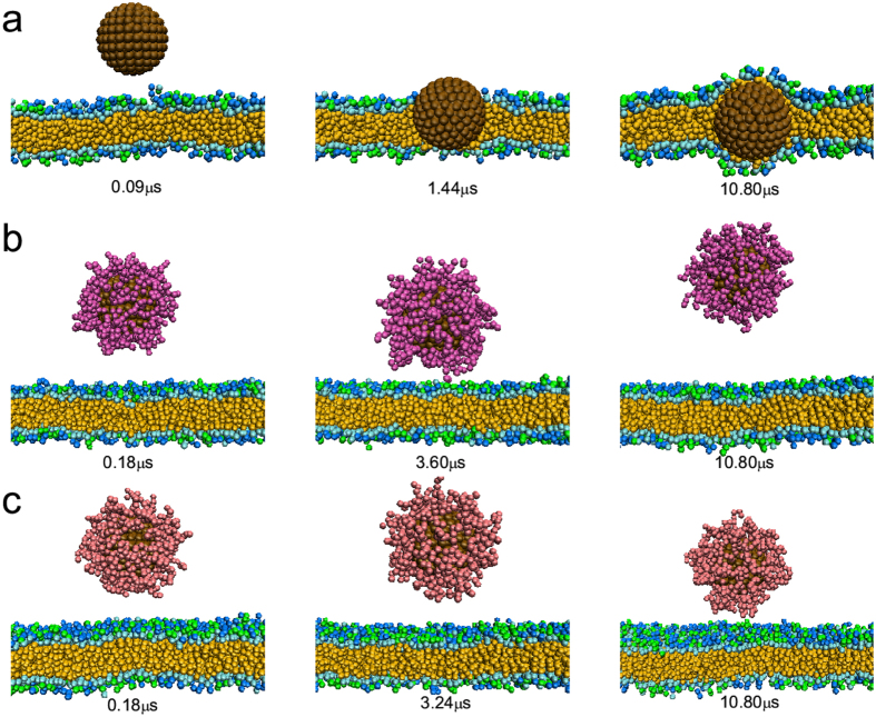
Figure 4. Time sequence of the snapshots of interactions between hydrophobic nanoparticle with different surface coatings and cell membranes where the nanoparticle size is 6 nm.
(a) Bare nanoparticle; (b) nanoparticle decorated with hydrophilic polymers (N = 8, σ = 0.8/nm2); (c) nanoparticle decorated with zwitterionic polymers (N = 8, σ = 0.8/nm2).

