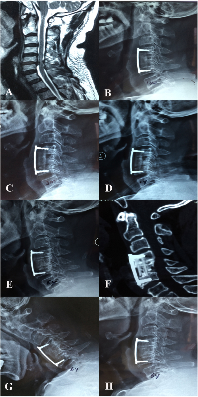
Figure 4. A 73-year old man who underwent 1-level ACCF with an n-HA/PA66 strut.
Preoperative MRI (A) scans revealed the lesion segment. A cervical lateral X-ray at one week after surgery (B) revealed that the strut was in the appropriate position, but the anterior titanium plate was too long. The n-HA/PA66 strut sank into the upper endplate, and internal fixation was dislodged at 3 months after surgery (C). At the 1-year follow-up, the subsidence of the n-HA/PA66 strut and the internal fixation migration were aggravated (D). A postoperative lateral X-ray (E) and 3D-CT (F) at the 6-year follow-up revealed bony fusion between the autograft inside the strut and the adjacent vertebrae. Osteophytes were also observed at the gap between the anterior plate and the upper/lower vertebral bodies. The flexion (G) and extension (H) radiographs revealed solid fusion in the fusion segment.

