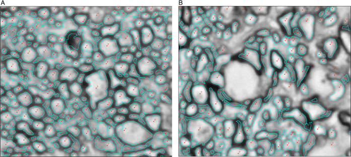
Figure 2. Examples of expert and AxonJ identified axons in PPD-stained 100x optic nerve sections.
(image set A, Table 1). Axon detections of expert (red point labels) and AxonJ (turquoise outlines), on a cropped 100x sample from image set A are depicted for the following: (A) one of the images where AxonJ performance was within 95% of the expert count, and (B) an image where AxonJ performance had only 88% correspondence to the expert.

