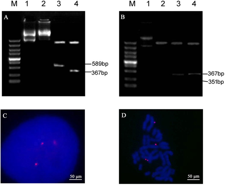
Figure 5. Determination of episomal vs. integrated status of plasmids in transfected CHO cells.
(A,B) DNA was extracted from CHO cells transfected with vectors containing the CMV and SV40 promoters, after which the extracted DNA was used to transform DH5α E. coli cells by electroporation. Transformants were selected and plasmid DNA was prepared and subjected to restriction analysis. Agarose gel electrophoresis of DNA extracted from CHO cells transfected with (A) the CMV promoter-containing vector and (B) the SV40-containing vector: M, DL5000; Lane 1, Extracted plasmid; 2, Kpn I digestion; 3, Ase I/Nhe I digestion; 4, KpnI/BamHI digestion. (C,D) Association of the episome with metaphase chromosomes from CHO cells transfected with vectors containing the CMV promoters. CHO cells transfected with plasmid were analysed by DNA FISH to assess whether the vectors were present as integrated copies. The episome (red) was visualized by eGFP FISH. Vector molecule distribution was monitored in post-mitotic nuclei of dividing cells (C) and episome localization was determined by FISH on spreads of metaphase chromosomes (D).

