
Fig. 7.
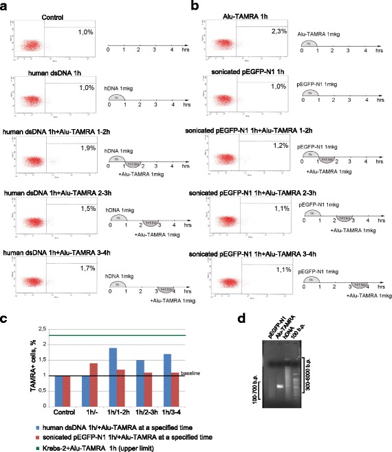
Competition between Alu-TAMRA DNA and high-molecular weight human DNA or sonicated pEGFP-N1 plasmid for internalization into Krebs-2 cells. a Addition of Alu-TAMRA DNA to Krebs-2 cells pre-incubated with human dsDNA for 1 h; b addition of Alu-TAMRA DNA to Krebs-2 cells pre-incubated sonicated pEGFP-N1 plasmid DNA. FACS plots and schematics of the experimental points analyzed are shown; c bar plot summarizing the percentages of TAMRA+ cells. Baseline equals background fluorescence of untreated cells; control represents cells incubated for 1 h with 1 μg of either human dsDNA (blue) or sonicated pEGFP-N1 plasmid (red); upper limit represents percentage of TAMRA+ cells after 1 h incubation with 1 μg Alu-TAMRA DNA; d results of an agarose gel electrophoresis showing fragment size distributions of the three eDNAs tested. Brackets on the sides of the gel indicate average sizes of sonicated pEGFP-N1 (left) and human dsDNA (right) fragments. dsDNA double-stranded DNA