
Fig. 6.
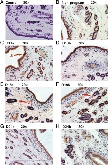
Immunhistochemical localization of HAND2 in pig uterus. GE = glandular epithelium; LE = luminal epithelium; S = stroma. a Negative control; b Immunohistochemical staining of non-pregnanct sows uterus with HAND2 antibody; c Immunohistochemical staining of porcine uterus attachment site with HAND2 antibody on d 13 of pregnancy; d Immunohistochemical staining of porcine uterus inter-site with HAND2 antibody on d 13 of pregnancy; e Immunohistochemical staining of porcine uterus attachment site with HAND2 antibody on d 18 of pregnancy; f Immunohistochemical staining of porcine uterus inter-site with HAND2 antibody on d 18 of pregnancy; g Immunohistochemical staining of porcine uterus attachment site with HAND2 antibody on d 24 of pregnancy; h Immunohistochemical staining of porcine uterus inter-site with HAND2 antibody on d 24 of pregnancy