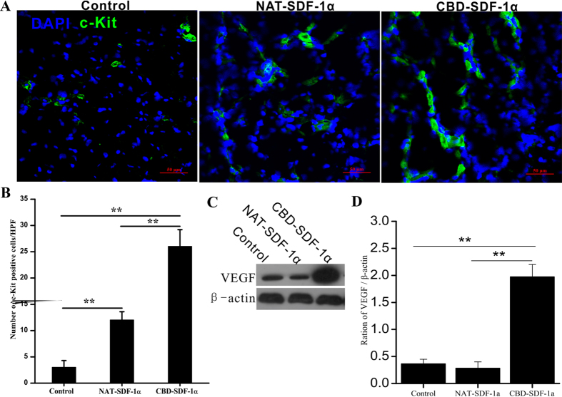
Figure 5. Recruitment of c-kit+ stem cells and the level of VEGF the ischemic area.
(A) Representative images of the c-kit+ stem cells that migrated into the ischemic area at 4 days after surgery. (B) Average number of c-kit+ cells in the infarcted heart (n = 6 in each group). CBD-SDF-1α mobilized more c-kit+ cells to migrate to the infarcted heart. (C) The level of VEGF in the ischemic area was detected by Western blot. (D) Quantification of the protein bands (n = 5 in each group). The data are presented as the means ± SEM, **P < 0.01.

