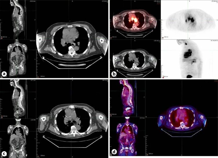
Fig. 1.

a CT scan with contrast demonstrating a large saddle filling defect. b PET-CT scan of the chest showing intense uptake in the mediastinum, hilar lymph nodes, and right thyroid. c Repeat CT scan of the chest with and without contrast after six courses of chemotherapy. d Repeat PET-CT scan showing resolution of intense FDG uptake after six courses of chemotherapy.
