Fig. 2.

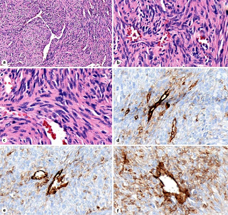
a H&E staining at low magnifiation. b, c H&E staining at higher magnification. d–f CD31 IHC stain showing a blood vessel (strongly CD31 positive) surrounded by weakly staining sarcoma cells indicative of vascular differentiation.

a H&E staining at low magnifiation. b, c H&E staining at higher magnification. d–f CD31 IHC stain showing a blood vessel (strongly CD31 positive) surrounded by weakly staining sarcoma cells indicative of vascular differentiation.