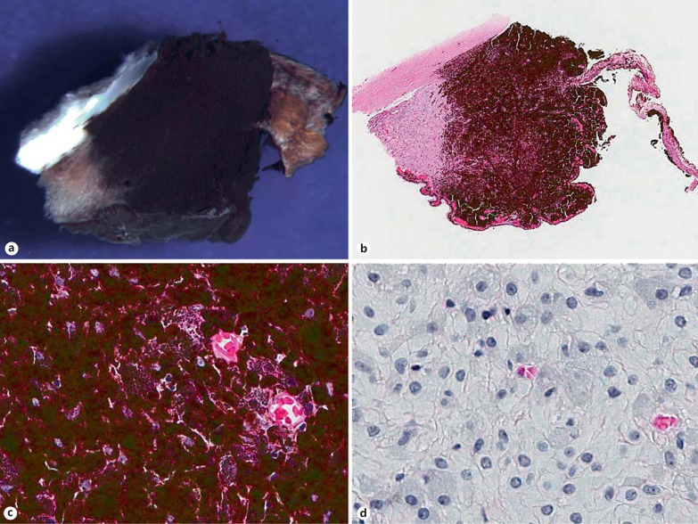
Fig. 1.
Iridociliary melanocytoma (specimen 1). Gross photograph (a) and low-power photomicrograph (b) of an iridocyclectomy specimen, demonstrating a darkly pigmented tumor, which involves the iris root and ciliary body stroma and extends into the anterior chamber angle (b HE stain; original magnification ×5). c The neoplasm is composed of polyhedral cells with dense intracytoplasmic pigments obscuring nuclear details (HE stain; original magnification ×100). d Bleached preparations highlight the bland nuclei with inconspicuous nucleoli, low nucleus-to-cytoplasm ratios and the absence of appreciable pleomorphism or mitotic figures (bleach-hematoxylin stain; original magnification ×100).

