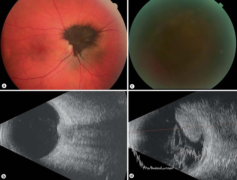
Fig. 2.
Optic nerve melanocytoma transformed to melanoma (specimen 6). Color fundus photograph of an optic-nerve melanocytoma at presentation (a) and the corresponding ultrasound image, demonstrating a 2-mm-thick tumor (b). The patient had a vision of 20/25 at that time. The color photograph at 3 years of follow-up demonstrates that the fundus is obscured by vitreous hemorrhage (c), at which point the patient's vision had decreased to light perception. The corresponding ultrasound (d) at that point demonstrates growth of the lesion to a height of 5.5 mm and overlying vitreous hemorrhage.

