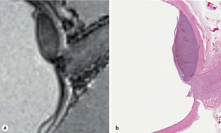
Fig. 2.
Ex vivo imaging. 7-tesla MRI of the same globe after enucleation. Prelaminar optic nerve extension is detectable (a). Photomicrograph of a pupil-optic nerve section with comparable axial orientation. Choroidal melanoma is seen impinging on the prelaminar optic nerve (b; HE, original magnification ×40).

