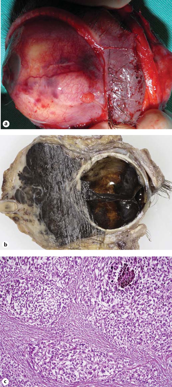
Fig. 3.
Exenteration specimen. a Gross view showing the subconjunctival Ahmed reservoir. b Sagittal cut demonstrates total retinal detachment, anterior advancement of the tumor and the massive retrobulbar tumor. c Histopathological analysis of the orbital part of the tumor reveals large epithelioid cells (HE, original magnification ×80).

