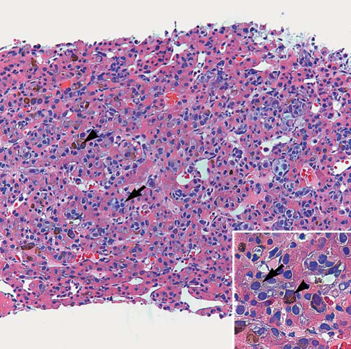
Fig. 3.
Metastatic melanoma infiltrates in the sinusoids of the liver. Residual hepatocytes with pink cytoplasm appear atrophic in-between the abundant melanoma cells (HE, ×200). Inset The neoplastic cells are plasmacytoid with light grey-purple cytoplasm and prominent nucleoli. Scattered melanoma cells exhibit cytoplasmic granular melanin pigment (HE, ×400). Arrows point to two (of numerous) nonpigmented melanoma cells, and arrowheads point to two pigmented melanoma cells.

