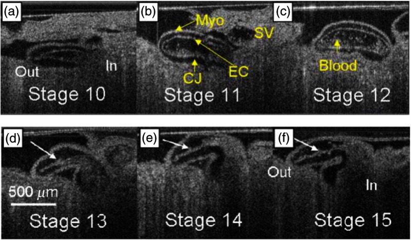
Fig. 19.
Time series of coronal sections at the same location of two quail hearts. (a–c) Stage 10 to 12 from the same quail embryo (Video 3, MPEG, 2.29 MB) [URL: http://dx.doi.org/10.1117/1.JBO.21.5.050902.3]. (d–f) Stage 13 to 15 from a different quail embryo, same orientation and location (Video 4, MPEG, 2.42 MB) [URL: http://dx.doi.org/10.1117/1.JBO.21.5.050902.4]. Adapted from Ref. 135.

