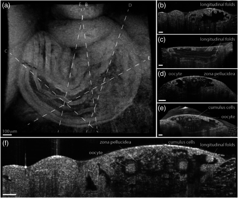
Fig. 22.
In vivo OCT imaging of the oviduct ampulla containing the oocytes and cumulus cells. (a) 3-D OCT reconstruction of the ampulla where the longitudinal mucosa folds are clearly visible. The dashed lines depict the cross sections shown in (b–f). All scale bars correspond to . Adapted from Ref. 161.

