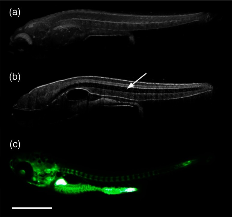
Fig. 8.
(a) Sagittal view of an OCT 3-D reconstruction of a 21 dpf wild type zebrafish in vivo. (b) 2-D cross-section where the spine is indicated by the white arrow. (c) Calcein stained image of the same specimen, taken an hour later. Scale bar is 1 mm. Adapted from Ref. 93.

