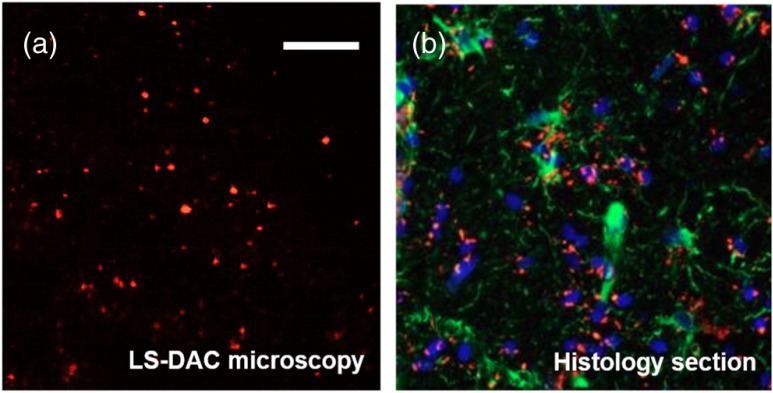
Fig. 5.
(a) Surgically resected glioma (brain tumor) specimen fluorescently imaged at 15 fps over a FOV of . The patient was administered 5-aminolevulinic acid (5-ALA) prior to surgery. (b) A histological section of a similar glioma specimen from a patient administered with 5-ALA prior to surgery. The image shows DAPI-stained nuclei (blue), 5-ALA-induced protoporphyrin IX fluorescence (red), and the expression of glial fibrillary acidic protein (green). .

