
Figure 5.
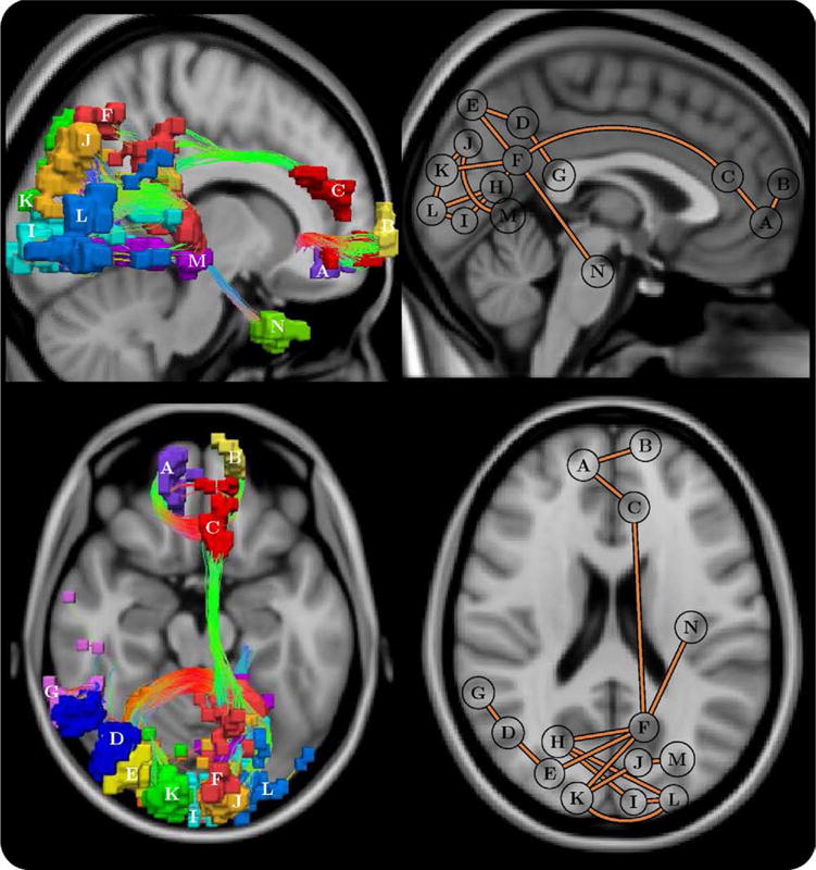
Schematic of the frontal-parietal/occipital network that was impaired in schizophrenia (p = .021 ± .004, corrected). Each connection comprising this network was impaired in patients but not in control subjects. Left: uniquely colored nodes and streamline representation of interconnecting fiber bundles. Anterior-posterior fibers: green; left–right: red; and superior-inferior: blue. Right: planar graph representation, where each node is depicted as a circle positioned at its node’s center of gravity. Note that the positioning of some posterior nodes was slightly shifted from the true center of gravity to avert overlapping. Top: sagittal, left hemisphere. Bottom: axial. Node abbreviations: (A) right medial orbital frontal, (B) left superior medial frontal, (C) left anterior cingulate, (D) right angular, (E) right superior occipital, (F) left precuneus, (G) right superior temporal, (H) right calcarine, (I) left calcarine, (J) left cuneus, (K) right cuneus, (L) left middle occipital, (M) left lingual, and (N) left fusiform.