
Fig. 5.
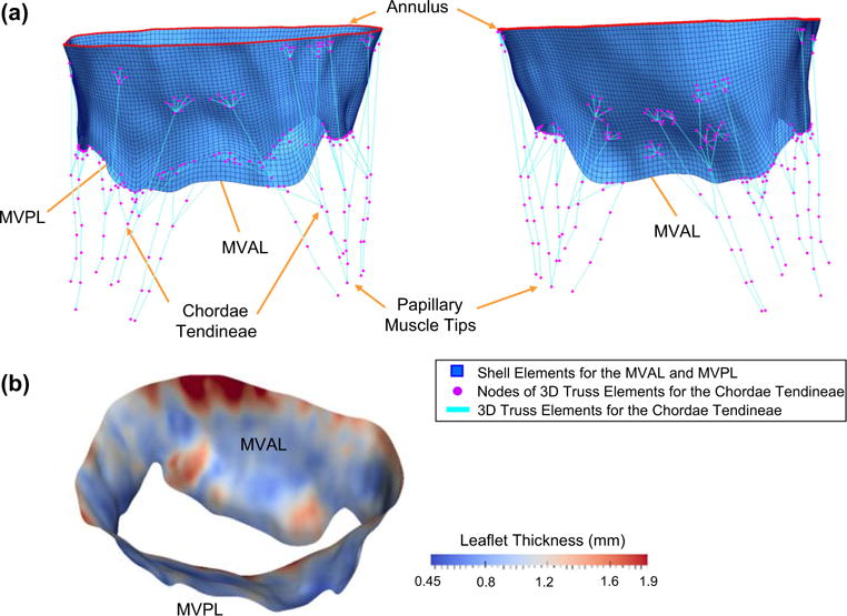
a Different views of the reconstructed FE model for the ovine MV apparatus with anatomically accurate leaflets represented by shell elements (blue), flat annulus that mimics the in vitro experimental clamped condition (red) and idealized chordae tendineae represented by 3D truss elements (cyan) with identified landmark points as FE nodes of the truss elements (magenta); b spatially varied thicknesses for both MVAL and MVPL, determined from the extracted atrial and ventricular leaflet surfaces, as part of input data for FE simulations