
Fig. 6.
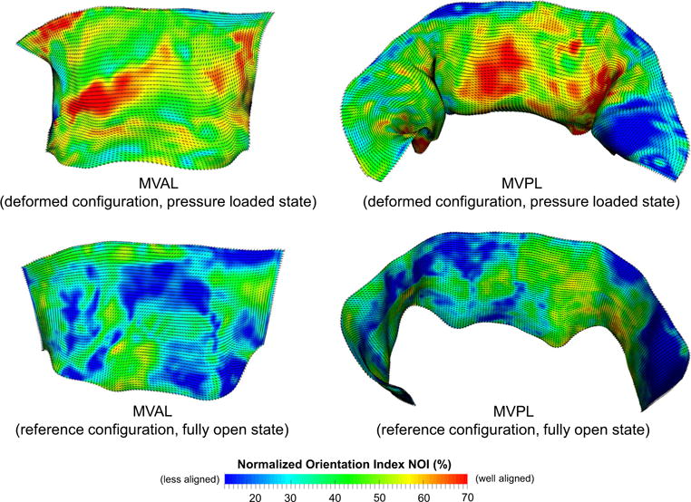
Results of the mapped fiber microstructural architecture for the MV FE mesh at the pressure-loaded state Ωt (top panel) and at the unloaded/reference state Ω0 (bottom panel). Dashed lines denote element-based local material axis direction and the color contour represents the strength of fiber splay. Note that the normalized orientation index is computed by NOI = (90° − OI)/90°