
Figure 1.
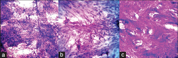
(a) Crush smear showing spindle cells of fibroblastic meningioma arranged in fascicles without meningothelial whorls, which was misinterpreted as schwannoma (H and E, ×200) (b) Crush smear of the same case showing the fascicular arrangement of spindle cells (H and E, ×400) (c) Frozen section of fibroblastic meningioma misinterpreted as schwannoma showing the spindle cell nature of the lesion due to thick sections (H and E, ×200)