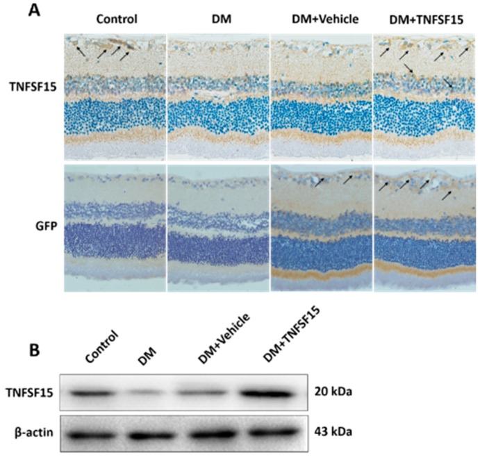
Figure 3.
Intravitreal injection of LV-TNFSF15-GFP successfully overexpressed TNFSF15 proteins in the retina of rats. (A) Typical images of TNFSF15 and GFP immunostaining in the retina. Arrows indicate TNFSF15 (upper panel) or GFP (lower panel) expression in the retina. Magnification, 400×. TNFSF15 was abundantly expressed in the retina of rats treated with LV-TNFSF15-GFP, but almost deficient in rats treated with vehicle (LV-NC-GFP). GFP was abundantly expressed in the retinas of rats treated with LV-TNFSF15-GFP and LV-NC-GFP, but absent in the control and DM group; (B) The TNFSF15 protein level was assessed by Western blot, and a representative image is shown.

