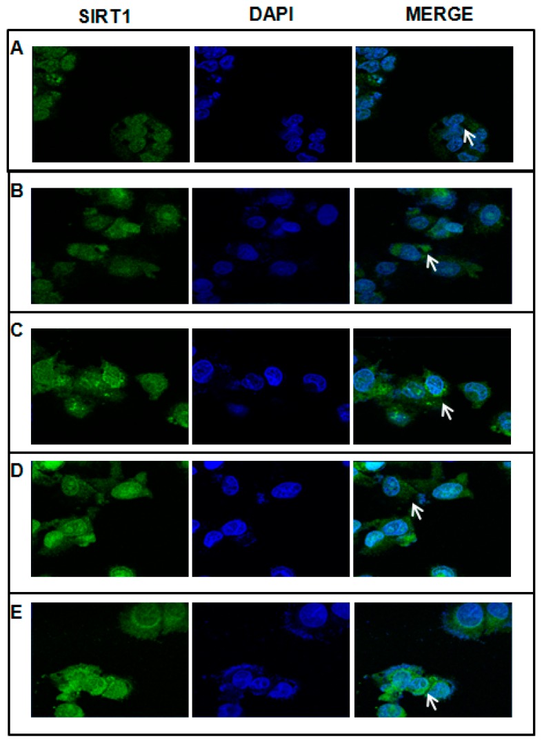
Figure 2.
Effect of AdoMet and Tyr on subcellular localization of SIRT1. HepG2 without treatment (A); Et-HepG2 (cells treated for 4 h with 1 M ethanol and then incubated for 48 h without ethanol) (B); Et-HepG2 treated for 48 h with 100 µM AdoMet (C); Et-HepG2 treated for 48 h with 10 µM Tyr (D); Et-HepG2 treated for 48 h with 100 µM AdoMet/10 µM Tyr (E). Cells were incubated with anti-SIRT1 (green). The nuclei were stained with DAPI (blue). The white arrows indicate the SIRT1localization. Images were obtained by an LSM-410 Zeiss confocal microscope.

