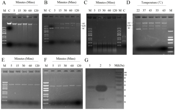
Figure 2.
Assay of rMbovNase nuclease activity. A–C: Different substrates, 1 µg BoMac cellular DNA (A); plasmid DNA (B); BoMac cellular RNA (C), incubated with 2.5 µg rMbovNase plus 10 mM CaCl2 in 100 mM Tris-HCl buffer (pH 8.5). Samples were collected at 5, 15, 30, 60, and 120 min and reactions were stopped by adding 10 mM EDTA and resolved on 1% agarose gels. Untreated DNA and RNA were negative controls (Lane C). Open-circle (OC), linear (LIN), and supercoiled (SC) forms of plasmid DNA are indicated; (D) rMbovNase had nuclease activity with plasmid DNA at temperatures from 22 to 65 °C. E: Nuclease activity of rMbovNaseΔ181–342. 2.5 µg rMbovNaseΔ181–342 (E) was incubated with plasmid DNA (1 μg) in the presence of 10 mM CaCl2 for 5, 15, 30, 60, or 120 min; PBS (F) was used as a negative control; (G) Zymogram analysis of nuclease activity using 12% SDS-PAGE gels containing 160 μg·mL−1 of herring sperm DNA in renaturation buffer. Lane 1, Total M. bovis cell lysate protein; Lane 2, Purified rMbovNase; Lane 3, Purified rMbovNaseΔ181–342.

